")
")
")


")

1. Atopic Eczema, Itch, and Pain
A case study presented at the 20th International Conference of the Society for Medical Innovation and Technology (SMIT) 2008 showed a reduction of itch for four hours and an overall reduction of itch from 8 to 3 (on a scale from 0 to 10) after daily CAP treatment for one minute of the left arm vs. basic treatment of the right arm over a period of 30 days. No side effects have been observed, and overall, the eczema of this patient was reduced by two points on a scale from -5 to +5.
However, a randomized two-sided placebo-controlled study on the efficacy and safety of atmospheric non-thermal argon plasma for pruritus with a total of 46 patients showed a similar improvement between plasma-treated and placebo-treated group with respect to itch. In this study, patients have been treated daily for two minutes with plasma or argon only (placebo). At the end of the study, a reduction of pruritus has been observed, which was likely due to standard therapy.
Another case study of a patient with chronic postoperative ear infection showed a highly significant reduction of pain after CAP application for local infection control. Considering these studies, cold atmospheric plasma seems to have positive effects on atopic eczema, itch, and pain, but still, more research is necessary to confirm these effects.
2. Disinfection (Bacteria/Fungi/Viruses)
Several studies have elucidated the lethality of CAP on bacteria and fungi and have shown its potential as an effective tool for disinfection. A two-minute CAP treatment, for example, has been shown to be effective against a variety of bacteria including important skin and wound pathogens such as Escherichia coli, group A Streptococcus, Methicillin-resistant Staphylococcus aureus (MRSA), and Pseudomonas aeruginosa, suggesting positive effects of CAP on wound healing.
Furthermore, the killing of clinically relevant fungal strains by CAP has been shown in vitro. A significant reduction of bacterial and fungal targets after plasma treatment has also been shown on model nails with onychomycosis (a fungal infection of the nail). The authors of this study conclude that the “CAP technology appears to be a safe, effective, and inexpensive therapy for fungal nail infection treatment.” Following this in vitro study, an in vivo pilot study evaluated the plasma treatment on 19 study participants with toenail onychomycosis.
No long-term sequelae have been observed after plasma treatment, and overall clinical cure was observed in 53.8% of participants, whereas mycological cure was observed in 15.4% of participants. A prospective randomized controlled study including 37 patients with herpes zosters (a painful skin infection caused by the varicella zoster virus) revealed that a weekday five-minute CAP treatment is safe, painless, and effective, improving initial healing of the herpes zoster lesions. Taken together, promising effects of CAP have been shown in regard to disinfection with no evidence for resistance of microorganisms against the treatment. Hence, CAP provides an effective method for skin disinfection.
3. Ichthyosis/Epidermal Barrier Defect
An acidic protective hydrolipid film produced by perspiratory glands and sebaceous glands covers the outer layer of the skin. This hydrolipid film provides an epidermal barrier that protects the skin from drying and contains a complex microbial ecosystem, consisting of numerous bacteria. The pH of the hydrolipid film is balanced between 5.4 and 5.9 in healthy skin.
Altered conditions in the hydrolipid film compared to healthy skin can lead to a shift in the microbial load and thus may promote disease. Pathogenic bacteria usually prefer pH values above 6; consequently, areas with increased pH values possess a higher susceptibility to pathogenic growth. The pH values in hydrolipid films of patients with diseases such as ichthyosis or atopic dermatitis have been shown to be higher compared to pH values of healthy skin.
One reason for an increased pH in patients with atopic dermatitis may be due to a mutation in filaggrin, a protein involved in the regulation of epidermal homeostasis. When human skin is treated with CAP, the hydrolipid film interacts directly with the chemical compounds of the plasma.
This prompted Helmke and colleagues to investigate the effect of CAP on pH of the hydrolipid film of diseased skin. Using a DBD plasma source, they treated lipid films of wool wax, pork sebum, and human lipid films with CAP and observed significant decreases in pH values. A treatment for only five seconds was sufficient to result in a decrease of pH, and treatment for 60 seconds led to a decrease of the pH down to 3.7 (from initial 4.6-6.2).
The pH values of wool wax after plasma treatment remained decreased for more than two hours. These studies provide first evidence for the potential treatment of epidermal barrier defects such as ichthyosis with CAP, where a decrease of the pH value would result in an inhibition of wound pathogens and, therefore, promote wound healing.
4. Wound Healing
In several clinical studies and case reports, the effect of CAP on wound healing has been assessed. Different plasma sources have been used in these studies (e.g., kINPen MED or PlasmaDerm VU-2010, Figure 2). The use of a hand-held dielectric barrier discharge plasma generator (PlasmaDerm® VU-2010) to alleviate chronic venous leg ulcers has been assessed in a monocentric, two-armed, open, prospective, randomized, and controlled trial.
This pilot study included 14 patients with at least one chronic venous ulcer, which had been divided into two comparable groups each consisting of seven patients. One group received standard care, while the other group in addition to standard care also received plasma therapy. Both groups were treated three times a week for a total of eight weeks with subsequent follow-up of four weeks.
While in both groups an ulcer size reduction of 50% has been observed, a greater size reduction (5.30 cm2 vs. 3.40 cm2) as well as a quicker size reduction after three weeks compared to the group without plasma treatment was found. Furthermore, there was one patient in the plasma-treated group who experienced complete healing of the ulcer. An example for successful wound healing after CAP treatment using the PlasmaDerm® VU-175 2010 is shown in Figure 3.
Further prospective randomized controlled studies using a different plasma device (MicroPlaSter predecessor of SteriPlas, Adtec Healthcare) primarily aimed at decreasing the bacterial load in chronic wounds. These studies including 36 and 24 patients, respectively, showed significant reductions of bacterial load in CAP-treated chronic wounds. A subsequent open retrospective study with 70 patients suggests that wound healing may be accelerated by CAP treatment.
Improved healing of pressure ulcers was also found in a prospective randomized controlled trial including 50 patients of which 25 received CAP treatment using a Bioplasma jet device. In another clinical case-control study, the efficacy of the plasma source kINPen MED was compared to octenidine treatment. This study including 16 patients with chronic leg ulcers revealed a similar reduction of bacteria using CAP treatment compared to octenidine.
A similar study with 34 patients showed a benefit of sequential treatment with CAP and octenidine dihydrochloride over treatment with just one of the two with respect to the antiseptic treatment. In addition to studies investigating the effect of CAP on chronic wounds, a few pilot studies also looked at the efficacy of CAP on acute wounds.
Heinlin and colleagues enrolled 40 patients with skin graft donor sites on the upper leg. Equally sized areas of the wounds were assigned randomly to receive either CAP or placebo treatment. From the second posttreatment day onwards, the CAP-treated sites showed significant improved healing compared to placebo treatment. In a case report study, four sterile ablative laser lesions (acute wounds) were induced in each of five volunteers. The wounds received either 10 seconds, three times 10 seconds, and 30 seconds CAP or no treatment for three consecutive days.
Treatment for three times 10 seconds and single treatment for 30 seconds showed best results in early stages of wound healing, and even at later stages (after six months and after one year), plasma treatment resulted in improved outcomes with respect to avoiding posttraumatic skin disorders.
Another study including six individuals with vacuum-generated wounds on the forearm analyzed wound healing parameters such as area decline and histomorphological characteristics. Wounds were treated with either no treatment, CAP, octenidine, or sequential treatment with CAP and octenidine. A statistically significant accelerated decline was observed after CAP treatment in comparison to the other treatment groups.
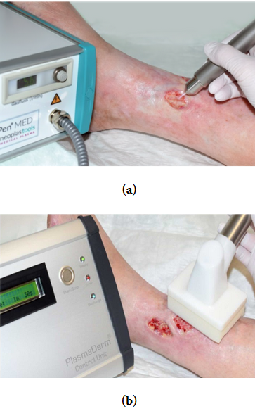
Figure 2: Treatment of chronic ulceration with cold atmospheric pressure plasma (CAP): (a) kINPen MED and (b) PlasmaDerm VU-2010.
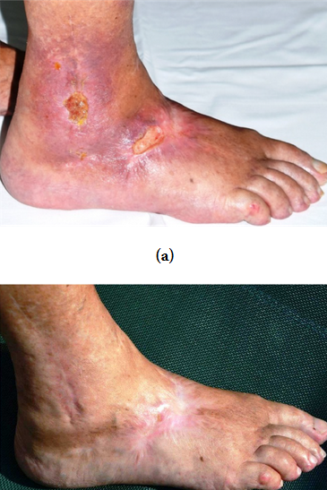
Figure 3: Example of successful wound healing after treatment with cold atmospheric pressure plasma (CAP): (a) chronic ulceration before CAP treatment and (b) complete healing for the first time since 14 years after CAP treatment for about 5 months.
Accelerated wound healing has also been observed in a mouse model treated with a cold plasma jet. In cell culture experiments with HaCaT keratinocytes and MRC5 fibroblasts, an increased motility of the cells has been observed after plasma treatment, and at least in HaCaT cells, this was associated with a decreased mRNA and protein expression of Cx43. Cx43 is a gap junctional protein of keratinocytes and was shown to inhibit cell migration and wound healing. These findings suggest that cold plasma enhances wound healing in chronic and slowly healing wounds.
There are several aspects of CAP that may contribute to an improved wound healing: UV radiation and reactive gas species (i.e., ozone) disinfect the wound, generation of nitric oxide (NO) or nitrogen species (NOx) stimulates the regeneration of tissue, and electric current stimulates angiogenesis. Furthermore, CAP leads to an acidification of the wound (decrease of pH).
5. Scar Treatment
The effect of CAP on tissue regeneration has also been elucidated for its potential in scar treatment. In one study, ten patients with acne scars received a single CAP treatment using a Plasma Skin Regeneration (PSR) system. According to patient and doctoral assessment, acne scars improved in 30% of the patients.
Thermal damage to the epidermis and upper dermis had been observed for four-six days, and besides that, collagen remodeling effects without permanent pigmentary or textural irregularities as well as regenerative epidermal effects have been described. Similarly, antimicrobiocidal effects of CAP and improved acne symptoms have been demonstrated using a direct DBD device. A reduction of P. acnes by 75% has been observed in a study with 31 volunteers.
Another cohort of 30 patients received weekly application of CAP for four weeks. This treatment led to a reduction of sebum production of 80%, which lasted four weeks following the treatment. Although the authors concluded that their plasma source can offer a new therapeutic option for acne treatment, more studies are needed to validate these findings.
6. Skin Tumors
Potential applications of CAP in cancer therapy are currently explored by several research groups. The possibility of a paradigm shift in cancer therapy and the selectivity to ablate cancer cells (e.g., melanoma) while the corresponding normal cells remain unaffected have already been described in 2011 by Keidar and colleagues.
Detachment of SW900 cancer cells from the culture vessel after plasma treatment has been observed, whereas no detachment was observed when normal human bronchial epithelial (NHBE) cells were treated. Furthermore, one single in vivo treatment with CAP for two minutes of ten mice with subcutaneous bladder cancer tumors (SCaBER) showed an ablation of small tumors (about five millimeters in diameter) and a size reduction of larger tumors. While fully ablated tumors did not recur, larger tumors started to regrow but did not reach their original size even after three weeks.
Next, the in vivo efficacy of CAP in a murine melanoma model has been investigated. A single plasma treatment induced ablation of the tumor and decreased the growth rate markedly. This also resulted in an increased survival rate in the treatment group, with a median survival of 33.5 days compared to 24.5 days in the control group. Following this study, a number of other studies assessed the efficacy of CAP as a potential therapy for cancer treatment.
Daeschlein and colleagues compared the antitumor efficacy of CAP with electrochemotherapy (ECT) in a melanoma mouse model. A single CAP treatment led to a significant delay of tumor growth acceleration. However, this was less effective compared to ECT, whereas the combination of CAP and ECT had the strongest effect. In light of these findings, the authors concluded that cold plasma provides a potential alternative to ECT and may serve as a new option for palliative skin melanoma therapy, either alone or in combination with ECT.
A murine melanoma B16/F10 cell line has been used to elucidate the effect of CAP against melanoma cells in vitro, showing a loss of viability of almost 100% 48 hours after CAP treatment for three minutes. In addition, in vivo treatment with CAP of F10 cells in mice showed a decrease in growth of the tumors which was even comparable to the decrease of tumor growth achieved with chemotherapy.
In a clinical study with six patients suffering from locally advanced head and neck cancers, the efficacy and side effects of cold plasma treatment have been explored. Two patients experienced a strong response to the treatment, resulting in a clear tumor reduction. While the tumor in one of these patients started to regrow ending with exitus letalis, the other patient was still receiving treatment, aiming for total remission. No side effects have been observed in two patients, while four patients experienced fatigue and a dry mouth. At least five of the patients had a reduction of odor, most likely due to decontamination, and four had a reduced demand of pain medication. Out of the six patients, five passed away after one to twelve months, which was not due to CAP application.
Besides ROS/RNS, the authors discussed the role of myeloid cells and the immunogenic cell death model of cancer treatment as potential mechanisms of action of CAP.
The potential use of CAP for cancer treatment has also been assessed in vitro and in vivo in several other nonskin cancer entities including breast cancer, pancreatic carcinomas, glioblastoma, colorectal carcinoma, and neuroblastomas. In these studies, growth inhibition of different cell lines in culture as well as decreased tumor growth or a reduction of tumor volume has been observed in different mouse models after CAP treatment.
Although the molecular mechanisms for the efficacy of CAP on cancer cells are not fully understood, reactive oxygen species (ROS) and charged particles have been determined to be major contributors to plasma-induced cell death.
Taken together, these findings indicate a general efficacy of CAP against various cancer entities, suggesting plasma to be a potential new therapy against diverse cancerous diseases. However, to integrate plasma treatment into modern cancer therapy, further studies have to be conducted.
7. Actinic Keratosis
The precancerous actinic keratosis is a patch of thick, scaly, or crusty skin. There are several options to treat actinic keratosis in order to prevent the development of squamous cell carcinomas. One option is the use of ingenol mebutate, which works in two ways: first, it leads to rapid lesion necrosis beginning one to two hours after application of ingenol mebutate, resulting from mitochondrial swelling and membrane disruption as well as membrane depolarization.
This process causes an inflammatory response, which, subsequently, leads to specific neutrophil-mediated antibody-dependent cellular cytotoxicity (ADCC) within days. These specific antibodies bind to specific antigens on dysplastic epidermal cells, as well as to receptors of infiltrating neutrophils, thereby causing the release of cytotoxic agents (e.g., ROS), which then destroy dysplastic epidermal cells.
The importance of the immune cell reaction against LK2 tumors (squamous cell carcinoma) has been shown in mice by Challacombe and colleagues. Following the treatment with PEP005 (ingenol-3-angelate), neutrophil-depleted mice showed a significant higher tumor volume in comparison to the control mice.
Similar to that, CAP seems to positively influence the healing of actinic keratosis as well. This has been shown in a pilot study with seven patients, who were treated twice a week, with CAP for two minutes, for a total of seven treatments. All patients have shown promising responses with no adverse events so that further investment does not only seem necessary but also very promising. In a retrospective study of 12 patients with advanced head and neck squamous cell carcinomas, CAP was used to decontaminate infected cancer ulcerations and evaluated for its anticancer effects.
Analysis of data from this study showed a decreased request for pain medication, a reduction of typical fetid odor related to a reduction of microbial load, and in some cases even a superficial partial remission of tumor and wound healing of infected ulcerations.
A case report from Daeschlein and colleagues showed promising results of CAP in a patient with recalcitrant actinic keratosis of the scalp. No actinic lesion relapse has been observed until at least 26 months after a single treatment with CAP, and furthermore, scar formation had been proven.
Taken together, these studies indicate a promising application of CAP as an innovative therapy for skin cancers or precancerous conditions, but the evidence is still sparse, and more basic research as well as more clinical studies are needed.
8. Conclusions
CAP has been shown to be a promising and inexpensive treatment for a variety of different diseases. While CAP already reached standard medical care status for wound treatment, only preliminary data for its effects in oncology are available. Continuing efforts in this emerging and highly dynamic field of plasma medicine will be necessary to further explore the full therapeutic potential of CAP and to fully understand its mechanisms of action. Whereas the development of novel plasma sources and modifications of existing devices will open up more opportunities, it will be crucial to adhere to certain standards in order to enable comparability of results obtained in different studies. Overall, the exciting field of plasma medicine is expanding and thus is providing increasing evidence for the use of CAP as a treatment option for a variety of dermatological diseases.
Source: https://www.hindawi.com/journals/omcl/2019/3873928/ref/#null